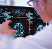
 Contributed by Center for Diagnostic Imaging Brain injuries are complex and can impair a person's physical and emotional functioning. They can be traumatic, resulting from accidents, falls or contact sports; or non-traumatic, resulting from strokes or aneurysms. Severe brain injuries can be difficult to treat. For all types of brain injuries, advanced imaging tools and expertise aids in accurate diagnosis and guiding a care plan. Diagnosis and Treatment Planning The treatment of head injuries depends on the type of injury and the severity. To assess the severity of a head injury, a physician may perform a physical and neurologic exam and imaging tests such as: COMPUTED TOMOGRAPHY (CT) A CT of the head and brain is often used as a first imaging test when a concussion is suspected. It is useful for detecting bleeding, swelling, brain injury and skull fractures.  MAGNETIC RESONANCE IMAGING (MRI) An MRI is helpful in detecting small hemorrhages or bruises, and monitoring changes in brain structure and function. It is especially useful in treatment planning.
VOLUMETRIC BRAIN IMAGING
This is performed with MRI post-processing software that provides objective, quantitative volume measurements of two conditions that can result from a brain injury: hydrocephalus and atrophy of the hippocampus. SUSCEPTIBILITY-WEIGHTED IMAGING (SWI) SWI is an MRI protocol run at a high resolution, increasing the ability to detect subtler injuries in patients with concussions/traumatic brain injuries, hemorrhages, neurodegenerative diseases and a variety of lesions. Learn about CDI’s outpatient medical imaging centers throughout Puget Sound and their specialized clinical teams and services at myCDI.com.
0 Comments
Your comment will be posted after it is approved.
Leave a Reply. |
Raise the BarRace reports, upcoming events, news, and more, from RTB. Archives
September 2023
|

 RSS Feed


